Call Us: (011) 41602514 / (011) 41602515 / (011) 41602516
How The Eye Works?
 Sight is an amazing
process made possible by many parts of the eye working
together. Light enters the eye and is bent or refracted by the
cornea (the window of the eye) and then passes through the
pupil (the opening in the iris). This light then passes
through the natural lens (located behind the pupil). This
completes the image formation by fine tuning the focused light
onto the retina. The retina changes the light (energy) into
electric impulses that are carried through the optic nerve to
the vision center (occipital cortex) of the brain where the
image is interpreted.
Cornea - The
cornea is the "window" of the eye (like a watch crystal). It
is the clear transparent part of the eye, through which the
colored part of the eye is seen. This is where the light is
bent the most and thus is the main source of refraction. The
cornea is made up of five layers of strong clear tissue,
without any blood vessels.
Sight is an amazing
process made possible by many parts of the eye working
together. Light enters the eye and is bent or refracted by the
cornea (the window of the eye) and then passes through the
pupil (the opening in the iris). This light then passes
through the natural lens (located behind the pupil). This
completes the image formation by fine tuning the focused light
onto the retina. The retina changes the light (energy) into
electric impulses that are carried through the optic nerve to
the vision center (occipital cortex) of the brain where the
image is interpreted.
Cornea - The
cornea is the "window" of the eye (like a watch crystal). It
is the clear transparent part of the eye, through which the
colored part of the eye is seen. This is where the light is
bent the most and thus is the main source of refraction. The
cornea is made up of five layers of strong clear tissue,
without any blood vessels.
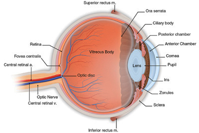
Sclera -The outer opaque "white part" of an eye is the sclera. This tough structure is the outer wall of the eye that gives protection to the delicate inner structures.
Choroid - This structure, between the sclera and the retina, is made up of blood vessels that provide nourishment to the eye.
Iris - This colored part of the eye has very fine muscles to control the size of the pupil.
Pupil - The pupil is the black-appearing spot in the center of the iris. Its size changes since its function is to control the amount of light reaching the retina. In the dark, it expands allowing more light to enter. It contracts in bright light to keep out excess light.
Lens - Located just behind the pupil it allows for changing of focus from distance to near objects by altering its shape. This changing focus is called accommodation. As a person ages the lens hardens and accommodation becomes more difficult.
Zonules - These are thread like structure which attach the lens to the ciliary muscle and help the lens to change its curvature during accomodation.
Ciliary Body - This contains two main structures. The first is a muscle that contracts and expands to control the curvature of the lens during accommodation. The second is a gland that secretes aqueous humor.
Aqueous Humor - This fluid is produced by the ciliary body and circulates in the front part of the eye. It provides nourishment to the front parts of the eye and maintains the eye pressure.
Retina - This membrane lines the inside wall of the eye. It contains photoreceptors (rods and cones) that change light into sight by converting light into electrical impulses. These electrical messages are sent from the retina to the brain and interpreted as images.
Macula - This tiny part of the retina is the central focusing spot. It is responsible for seeing details (such as reading) and also for color vision.
Optic Nerve - This nerve is the pathway that the light rays take from the retina to the processing center of the brain. It actually is made of about a million tiny nerves bundled together.
Optic Disc - This area is not sensitive to light and it is often referred to as the "blind spot". It is where the retina meets the optic nerve.
Vitreous Gel - This clear gel fills the central core of the eye. It helps to maintain a spherical shape to the eye.
Sight is an amazing
process made possible by many parts of the eye working
together. Light enters the eye and is bent or refracted by the
cornea (the window of the eye) and then passes through the
pupil (the opening in the iris). This light then passes
through the natural lens (located behind the pupil). This
completes the image formation by fine tuning the focused light
onto the retina. The retina changes the light (energy) into
electric impulses that are carried through the optic nerve to
the vision center (occipital cortex) of the brain where the
image is interpreted.
Cornea - The
cornea is the "window" of the eye (like a watch crystal). It
is the clear transparent part of the eye, through which the
colored part of the eye is seen. This is where the light is
bent the most and thus is the main source of refraction. The
cornea is made up of five layers of strong clear tissue,
without any blood vessels. Sclera -The outer opaque "white part" of an eye is the sclera. This tough structure is the outer wall of the eye that gives protection to the delicate inner structures.
Choroid - This structure, between the sclera and the retina, is made up of blood vessels that provide nourishment to the eye.
Iris - This colored part of the eye has very fine muscles to control the size of the pupil.
Pupil - The pupil is the black-appearing spot in the center of the iris. Its size changes since its function is to control the amount of light reaching the retina. In the dark, it expands allowing more light to enter. It contracts in bright light to keep out excess light.
Lens - Located just behind the pupil it allows for changing of focus from distance to near objects by altering its shape. This changing focus is called accommodation. As a person ages the lens hardens and accommodation becomes more difficult.
Zonules - These are thread like structure which attach the lens to the ciliary muscle and help the lens to change its curvature during accomodation.
Ciliary Body - This contains two main structures. The first is a muscle that contracts and expands to control the curvature of the lens during accommodation. The second is a gland that secretes aqueous humor.
Aqueous Humor - This fluid is produced by the ciliary body and circulates in the front part of the eye. It provides nourishment to the front parts of the eye and maintains the eye pressure.
Retina - This membrane lines the inside wall of the eye. It contains photoreceptors (rods and cones) that change light into sight by converting light into electrical impulses. These electrical messages are sent from the retina to the brain and interpreted as images.
Macula - This tiny part of the retina is the central focusing spot. It is responsible for seeing details (such as reading) and also for color vision.
Optic Nerve - This nerve is the pathway that the light rays take from the retina to the processing center of the brain. It actually is made of about a million tiny nerves bundled together.
Optic Disc - This area is not sensitive to light and it is often referred to as the "blind spot". It is where the retina meets the optic nerve.
Vitreous Gel - This clear gel fills the central core of the eye. It helps to maintain a spherical shape to the eye.
About Our Special Facilities


Facilities & Recognition
1) Registered Nursing
2) Organ (Cornea)
Transplant Centre
2) Organ (Cornea)
Transplant Centre

Executive Eye Checkup
Many of the eye problems can be prevented, controlled and cured if detected at an early stage.